Keeping you informed with the latest 
news, research, new products and events.
Showing posts with label electron microscopy. Show all posts

A new blog for Microscopy and Analysis

Posted 3/03/2011 5 comments
I have a new blog I'm writing for Microscopy and Analysis. Every few weeks I'll sample some of the biggest news and research in microscopy and put it into a Microscopy News Round Up post.

Here are some of my recent posts:

Happy Reading!

HD comes to electron microscopy

Posted 7/18/2010 0 comments
Carl Zeiss has introduced the EVO HD conventional scanning electron microscope (SEM). The system delivers higher resolution at low acceleration voltages compared to present conventional SEM and bring High Definition to electron microscopy.

The new EVO HD source has a higher brightness, which offers a better resolution at low-kV than conventional tungsten SEMs. The source also aids analytical applications, giving a 30% increase in resolution at 30kV and 1nA.

The company says that numerous applications in both life sciences and materials analysis will benefit from the increased performance of the microscope.
Caption: This image of a scale from butterfly wing (Pieris Brassicae) was taken on the EVO HD electron microscope at 5kV acceleration voltage.

More information about the microscope here.

Arizona State University Dedicates Room in Honor of a Microscopy Lab Manager

Posted 7/05/2010 0 comments
The John Wheatley Education and Outreach Room was recently dedicated at Arizona State University’s LeRoy Eyring Center for Solid State Science. Wheatley managed the center’s John M. Cowley Center for High Resolution Electron Microscopy lab for 25 years before his death in 2005. He was responsible for operations of what is one of the nation’s largest collections of electron microscopes. He is credited by colleagues for his contributions to making it one of the leading electron microscopy facilities in the world.

The Wheatley Room will be used for Science is Fun, an education outreach program for middle school and high school students, as well as for training and education of university students, for teachers from K-12 schools and colleges, and for use by university faculty and industry researchers.

Eyring Center professor and researcher Ray Carpenter said Wheatley established a tradition of excellence through his day-to-day management of the lab. “Electron microscopes are complex electromechanical instruments that require constant attention and care to operate at peak performance levels. John Wheatley was remarkably successful at this task,” Carpenter said.

The lab’s history of success under Wheatley’s watch, he said, was a significant factor in gaining support from the National Science Foundation in recent years for the center to acquire state-of-the-art aberration-corrected electron microscopes, due to be installed in 2011.

McCrone to distribute JEOL NeoScope

Posted 5/17/2010 0 comments
McCrone Microscopes & Accessories is now a licensed distributor of the JEOL NeoScope, a benchtop scanning electron microscope (SEM). McCrone Microscopes & Accessories is the instrument sales division of The McCrone Group.

The JEOL NeoScope is a compact, automated SEM designed for analysis of biological and material samples. Unlike other SEMs, commonly used settings are automated with the NeoScope, making the instrument simple to use without sacrificing image resolution. The microscope can be used to supplement optical microscopic analysis for a wide variety of applications, including pharmaceutical, geological and forensics studies. he high resolution, large depth of field and automated settings (focus, brightness and contrast) make the unit easy-to-use and accessible to those in classroom settings or research laboratories, as well as novice or advanced SEM users. In addition, no special sample preparation is needed for conductive and non-conductive samples, further simplifying the process for the user.

On-site training, service contracts and a full line of SEM accessories for the NeoScope are available through McCrone Microscopes & Accessories.

Researchers image virus structure at atomic resolution

Posted 5/04/2010 0 comments
UCLA researchers led by Hong Zhou have used cryo-electron microscopy to image a virus structure at 3.3 angstroms. Structurally accurate 3-D reconstructions of biological complexes are possible with cryo-electron microscopy because the samples are flash frozen, which allows them to be imaged in their native environment. Also, the microscope operates in a vacuum an environment better for electron travel.

The researchers studied the structure of the aquareovirus, a non-envelope virus that causes disease in fish and shellfish, in an effort to better understand how non-envelope viruses infect host cells. Envelope viruses such as influenza and HIV have an envelope-like membrane that the virus uses to fuse with and infect a host cell. Non-envelope viruses lack this membrane and infect cells with a poorly understood process that involves using a protein to fuse with the cells.

The high-resolution 3-D cryo-electron microscopy images showed that the aquareovirus uses a priming stage to accomplish cell infection. In its dormant state, the virus has a protective protein covering, which it sheds during priming. Once primed, it is ready to use a protein called an "insertion finger" to infect a cell.

Zhou said that better knowledge of virus structures could help with engineering of medications that work against viruses or that use engineered viruses to deliver drugs and gene therapy.
Cell, Volume 141, Issue 3, 472-482, 15 April 2010 DOI: 10.1016/j.cell.2010.03.041

New microscopy laboratory to focus on nanotechnology

Posted 5/03/2010 0 comments
Trinity College Dublin's Centre for Research on Adaptive Nanostructures and Nanodevices (CRANN) opened its Advanced Microscopy Laboratory last week. The laboratory has advanced instrumentation for viewing material on the atomic scale and was funded by the Higher Education Authority and Science Foundation Ireland. It was officially opened by the Minister for Labour Affairs, and Public Service Transformation, Mr Dara Calleary, TD (pictured with CRANN researcher Dr. Despina Bazou).
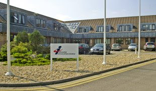
The €12-million microscopy laboratory houses a helium ion microscope, a scanning electron microscopes, and a focused ion beam. The helium ion microscope allows the highest resolution surface imaging of bulk materials from a scanning microscope and is the only such tool installed in Ireland.

More information at http://www.crann.tcd.ie/index.

Late summer/fall microscopy events

Posted 8/11/2009 0 comments
August 19-21, 2009 
International Tutorial Workshop on Piezoresponse Force Microscopy (PFM)
National Institute for Materials Science (NIMS)
Tsukuba, Japan
The workshop is the 5th in a series and precedes the IMF-ISAF conference at Xi'an Jiaotong University in China. It will feature a series of tutorial and invited lectures on instrumental and theoretical aspects of PFM, nanoscale ferroelectricity, and emerging techniques such as Scanning Nonlinear Dielectric Microscopy. More information here

August 26, 2009
Materials Electron Microscopy: At the Limit
University of Illinois at Chicago
USA
This Midwest Microscopy & Microanalysis Society Meeting is a day of invited speakers discussing advanced electron microscopy in materials science. More information here

September 8-11, 2009
EMAG Conference and Exhibition 
University of Sheffield
UK
The Electron Microscopy and Analysis Group’s (EMAG) biennial conference captures the latest in the development and applications of electron microscope techniques. This year's themes are advanced electron microscopy techniques; investigating structure-property relationships in advanced material; and nanophysics and nanotechnology. The conference includes exhibitors, lectures, posters and technical workshops. More information
here

September 22-25, 2009
Force Measurement and Manipulation in Biological Microscopy
University of North Carolina
Chapel Hill, NC USA
Learn the theory and techniques of measuring forces in biological samples in this course presented by Computer Integrated Systems for Microscopy and Manipulation, an NIH NIBIB Biotechnology Resource Center at The University of North Carolina at Chapel Hill. Attendees will use AFM, laser tweezers, and 3-D magnetic force systems on live biological samples, learn the theory and applications of force measurement, and receive an overview of mechanisms of force generation in biology and consequences at molecular and cellular level. More information here

October 14-15, 2009
8th International Symposium on Scanning Probe Microscopy & 
2nd International Symposium on Optical Tweezers in Life Sciences
Umspannwerk Ost, Berlin

This joint meeting is a forum for applications in scanning probe and optical tweezers technologies in life sciences. Applications covered include biophysical, biochemical and medical research. Leaders in the field together with novices are encouraged to learn from each other, discuss the presentations and share their experiences. More information here.

These events are also listed in the calendar on the right of this page. 

Day 1 at Microscopy and Microanalysis 2009

Posted 7/28/2009 0 comments
I arrived in Richmond, VA around noon today and checked in with the Microscopy and Analysis staff. I'm providing blog coverage of the conference for the M&A journal's web site. The staff was very nice and made sure I had everything I needed while giving me the space to do my own thing. I got the opportunity to use a Flip camera for a little video blogging, but decided to listen to talks instead today. 

The show seemed a little quiet, but I think it is just because it is a smaller niche show being in held in a rather large conference center. It has been a few years since I had been to this show and I had forgotten how centered on electron microscopy the exhibit floor is. This always struck me as strange since the research presented here is more evenly divided among microscopy techniques. 

I had planned to do some live twittering from the show, but was disappointed that the free wireless promised by the conference center web site was only in certain spots (actually, I only found one spot). This meant to even e-mail in my blog posts, I had to walk across the street. It wasn't that far, but it caused me to miss at least one talk and made it impossible to Tweet throughout the day. 

I think I'll have a better pace set for tomorrow. For today, I covered two presentations. Click on the descriptions below to read the full blog post.


Microscopy news briefs- Fulbright, Asylum Research, Genetix

Posted 5/21/2009 0 comments
Fulbright Scholarships awarded for microscopy research
Jonathan Winterstein, a doctoral student in materials science & engineering at the University of Connecticut has been awarded a Fulbright scholarship to perform research at the Technical University of Graz’s Austrian Centre for Electron Microscopy and Nanoanalysis. His research will focus on ceramic fuel cell materials, which have potential for use in clean energy generation. Winterstein plans to use transmission electron microscopy and spectroscopy to study defects in ceramic materials. Controlling these defects could optimize the performance of fuel cell materials.

Asylum Research UK moves to Bicester, Oxfordshire
On May 26th, Asylum Research’s wholly owned UK subsidiary will move to a larger office in Bicester, Oxfordshire. The additional space will give the company room for more staff and allow them to demo their Cypher AFM system, which will join the MFP-3D demo system.

Genetix acquires SlidePath Limited
The cell imaging and analysis company Genetix Group plc (New Milton, UK) has acquired all the issued share capital of the digital pathology company SlidePath Limited (Dublin). The acquisition will further strengthen Genetix’s position in the digital pathology market. For example, Genetix will add SlidePath’s web-enabled viewing, information management and computer-aided image analysis capabilities to its products.

Electron microscopy class projects

Posted 5/12/2009 0 comments
Students from Brian McIntyre's electron microscopy class at the University of Rochester had some great final projects. Here are just a few images from the projects.    

These images from Jonathan M. Malecki show a macrophage taking up multi-walled nanotubes. 

Amanda Carey studied airborne particulate matter emitted from a coal power plant. The image shows a  large iron-rich grain and a smaller spherical iron-enriched grain in the lower right. The smaller grain is magnified in the bottom image. 


Check out more of the projects, including 3D images, here

Transmission electron microscopy image of swine flu

Posted 4/29/2009 1 comments
Since there have been some funny microscopy images of swine flu and also some circulating pictures that weren't actually of swine flu, I wanted to be sure everyone could see the real thing. 

This colorized negative stained transmission electron micrograph (TEM) depicts some of the ultrastructural morphology of the A/CA/4/09 swine flu virus. Courtesy of CDC/ C. S. Goldsmith and A. Balish. 

New CMOS camera and workflow software

Posted 4/17/2009 0 comments

Capture color
If you’re looking for versatile yet affordable color camera, you might try the  Olympus SC30 microscopy camera that was recently introduced in Europe. Suitable for material and life science applications, it has a native resolution of 2048 x 1532 pixels, uses a 3.3 megapixel CMOS chip, features exposure times that can be adjusted from 57 µs to 1.75 s, and has binning modes of 2x, 3x, and 4x. It can be used for live cell imaging, standard bright field applications, and for digital documentation. With 4x binning, it can capture 49 fps at resolution of 508 x 384 pixels. More info here

Get more done
Macnification digital electron microscopy shareware for the Mac could increase your productivity. It helps users organize, find, annotate, analyze, adjust, compare, and publish microscopic images. All major image file formats can be imported, and measurement results can be exported to spreadsheet applications. Requires Mac OS X 10.5 or later. More info here

ACS Spring Meeting

Posted 3/24/2009 0 comments
The Spring '09 meeting of the American Chemical Society is currently underway in Salt Lake City. With the theme "Nanoscience: Challenges for the Future," there's sure to be some interesting microscopy research being presented.

Here are a few of my picks:
  • Studying spores with atomic force microscopy (AFM) – Alexander Malkin from the Lawrence Livermore National Laboratory discusses how in vitro AFM can be used to study microbial systems. For example, it can be used for directly visualizing the structural dynamics of single germinating bacterial spores. Yet another example of AFM's usefulness in biology.
  • Organic photovoltaics under the microscope – Researchers from the University of Texas at Austin simultaneously collected near-field scanning optical microscopy (NSOM) images of transmission, fluorescence, and photocurrent to examine factors limiting efficiency of an organic photovoltaic device. Organic photovoltaics are one of the leading technologies for solar power.
  • Mixing microscopy and food– Researchers at Auburn University have used AFM to study of the nanostructure of food grade gelatin and polysaccharide water absorbents as well as for rapid identification of microorganisms tied to food safety. With all the food safety problems in the news, techniques to make sure food is safe before it gets to consumers are needed.
  • MEMS-based heater for electron microscopy - Researchers from Oak Ridge National Laboratory together with Protochips Inc. are developing an in-situ MEMS-based heater chip that will aid in electron microscopy imaging of mononuclear catalytic species such as Au/Fe2O3 and Ir/MgO.

The conference included several talks on optical methods that overcome the diffraction limit:
Fluorescence photoactivation localization microscopy (FPALM)
Stochastic optical reconstruction microscopy (STORM)
Using fusions to EYFP
These all seem really nifty to me, but I've been hearing about various techniques to overcome the diffraction limit for several years (maybe more than several). What will it take for these techniques to find more use?
 

Microscopy News © 2008 using D'Bluez Theme Designed by Ipiet Supported by Tadpole's Notez Based on FREEmium theme Blogger Templates